Author: Dr Neha choudhary

  • How to Read Blood Test Reports

    How to Read Blood Test Reports

    Category: Clinical Skills
    Tags: Blood Tests, Laboratory Reports, CBC, Blood Test Interpretation, Medical Students, Healthcare Education, Clinical Skills

    Introduction

    Blood tests are among the most commonly used investigations in healthcare. They can provide valuable information about a person’s blood cells, organs, metabolism, immune system, and overall health.

    For medical and healthcare students, learning how to read a blood test report is an important clinical skill. However, interpreting laboratory results correctly requires more than simply checking whether a number is marked “high” or “low.”

    In this beginner-friendly guide, we will look at the common sections of a blood test report, understand what the major tests generally indicate, and learn how to approach laboratory results systematically.

    Important: Reference ranges vary between laboratories and may depend on age, sex, pregnancy, medications, medical history, and testing methods. A single abnormal result does not necessarily mean that a person has a disease. Clinical interpretation should be performed by a qualified healthcare professional.


    What Is a Blood Test?

    A blood test involves collecting a sample of blood and analyzing it in a laboratory.

    Depending on the type of test ordered, the sample can provide information about:

    • Blood cells
    • Blood glucose
    • Kidney function
    • Liver function
    • Electrolytes
    • Cholesterol
    • Inflammation
    • Hormones
    • Nutritional markers
    • Other disease-related markers

    A doctor may order one test or a combination of tests depending on the patient’s symptoms and clinical needs.


    Understanding a Blood Test Report

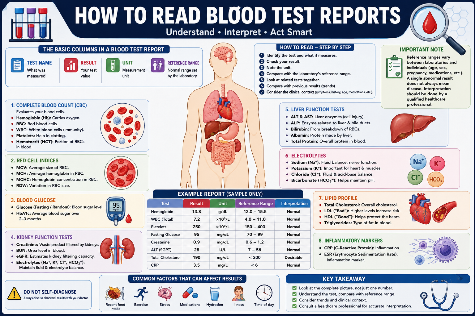
    A typical laboratory report may contain several columns.

    For example:

    TestResultUnitReference Range
    Hemoglobin13.8g/dLLab-specific
    WBC7.2×10⁹/LLab-specific
    Platelets250×10⁹/LLab-specific
    Glucose95mg/dLLab-specific

    The important information includes:

    Test Name

    This tells you what was measured.

    Result

    This is the value obtained from the patient’s blood sample.

    Unit

    Different tests use different measurement units.

    Reference Range

    This is the range the laboratory uses when interpreting the result.

    Always look at the reference range printed on the report, rather than relying on a range from another laboratory.


    1. Complete Blood Count (CBC)

    A Complete Blood Count (CBC) is one of the most common blood tests.

    It provides information about different types of blood cells.

    Major components include:

    • Red blood cells
    • White blood cells
    • Hemoglobin
    • Hematocrit
    • Platelets
    • Red-cell indices

    Hemoglobin

    Hemoglobin is a protein in red blood cells that carries oxygen.

    A lower-than-expected hemoglobin level may occur in conditions such as anemia, but the cause requires further evaluation.

    Possible causes of anemia include:

    • Iron deficiency
    • Vitamin deficiencies
    • Blood loss
    • Chronic diseases
    • Other medical conditions

    A healthcare professional usually considers hemoglobin together with other CBC values and the patient’s clinical history.


    Red Blood Cells

    Red blood cells (RBCs) carry oxygen from the lungs to tissues and help transport carbon dioxide back toward the lungs.

    The RBC count is interpreted together with hemoglobin, hematocrit, and red-cell indices.


    Hematocrit

    Hematocrit (HCT) represents the proportion of blood volume occupied by red blood cells.

    Changes in hematocrit can occur with conditions affecting red blood cells or blood volume.


    White Blood Cells

    White blood cells (WBCs) play an important role in the immune system.

    Changes in WBC count can occur with many different conditions, including infections, inflammation, medications, and disorders affecting blood cells.

    A WBC result should therefore not be interpreted as proof of infection by itself.


    Platelets

    Platelets help the blood form clots.

    A platelet count that is significantly outside the laboratory’s reference range may require further clinical evaluation.

    Both low and high platelet counts can have different causes.


    2. Red Blood Cell Indices

    CBC reports may include several red-cell indices.

    MCV

    Mean Corpuscular Volume (MCV) describes the average size of red blood cells.

    It can help healthcare professionals classify certain types of anemia.

    MCH

    Mean Corpuscular Hemoglobin (MCH) represents the average amount of hemoglobin in a red blood cell.

    MCHC

    Mean Corpuscular Hemoglobin Concentration (MCHC) reflects the concentration of hemoglobin within red blood cells.

    RDW

    Red Cell Distribution Width (RDW) describes variation in the size of red blood cells.

    These values are generally interpreted together rather than individually.


    3. Blood Glucose

    Blood glucose testing measures the amount of glucose (sugar) in the blood.

    Depending on the test, glucose may be measured:

    • While fasting
    • After eating
    • As part of a random blood glucose test
    • As part of longer-term glucose assessment

    HbA1c

    HbA1c, also called glycated hemoglobin, provides an estimate of average blood glucose over approximately the previous few months.

    It is commonly used in the assessment and monitoring of diabetes.

    The interpretation of glucose and HbA1c results depends on the testing context and the individual’s clinical situation.


    4. Kidney Function Tests

    Blood tests can provide information about kidney function.

    Common measurements include:

    • Creatinine
    • Blood urea nitrogen (BUN), where used
    • Estimated glomerular filtration rate (eGFR)
    • Electrolytes

    Creatinine

    Creatinine is a waste product that is removed from the blood by the kidneys.

    Changes in creatinine can occur for several reasons, so the result should be interpreted alongside other kidney-related measurements and the patient’s clinical history.

    eGFR

    Estimated glomerular filtration rate (eGFR) is an estimate of kidney filtration.

    It is commonly used to assess kidney function, but it is not a direct measurement and should be interpreted appropriately for the individual patient.


    5. Liver Function Tests

    Blood tests can also provide information about the liver.

    Commonly measured markers include:

    • ALT
    • AST
    • ALP
    • Bilirubin
    • Albumin
    • Total protein

    ALT and AST

    ALT and AST are enzymes that can increase when cells are injured.

    They are often used as part of liver evaluation, although they are not completely specific to the liver.

    Bilirubin

    Bilirubin is produced during the normal breakdown of red blood cells.

    Elevated bilirubin can occur for different reasons, including conditions affecting the liver, bile flow, or red blood cell breakdown.

    Albumin

    Albumin is a major protein produced by the liver.

    Albumin levels can be affected by several conditions and should be interpreted in context.


    6. Electrolytes

    Electrolytes are minerals that play important roles in fluid balance, nerve function, and muscle function.

    Commonly measured electrolytes include:

    • Sodium
    • Potassium
    • Chloride
    • Bicarbonate or total CO₂

    Sodium

    Sodium is important for maintaining fluid balance and normal nerve and muscle function.

    Potassium

    Potassium is particularly important for muscle and heart function.

    Significant abnormalities in potassium can require urgent medical attention depending on the level and clinical situation.


    7. Cholesterol and Lipid Profile

    A lipid profile may include measurements such as:

    • Total cholesterol
    • LDL cholesterol
    • HDL cholesterol
    • Triglycerides

    LDL

    LDL cholesterol is often described as a cholesterol component associated with increased cardiovascular risk when elevated.

    HDL

    HDL cholesterol participates in the transport of cholesterol and is one component considered when evaluating cardiovascular risk.

    Triglycerides

    Triglycerides are a type of fat found in the blood.

    Lipid results should be considered together with other cardiovascular risk factors rather than interpreted in isolation.


    8. Inflammatory Markers

    Some blood tests are used to assess inflammation.

    Two commonly known markers are:

    • CRP — C-reactive protein
    • ESR — erythrocyte sedimentation rate

    These tests can indicate that inflammation may be present, but they generally do not identify the specific cause by themselves.

    Further clinical evaluation may be necessary.


    High and Low Results: What Do They Mean?

    Many laboratory reports mark results as:

    • High
    • Low
    • Abnormal
    • Outside reference range

    However, an abnormal result does not automatically mean that a person has a disease.

    For example, a result can be influenced by:

    • Recent food intake
    • Exercise
    • Hydration
    • Medications
    • Age
    • Pregnancy
    • Time of day
    • Temporary illness
    • Laboratory methods

    This is why laboratory results should be interpreted alongside the patient’s symptoms, medical history, examination, and other investigations.


    How to Read a Blood Test Report Step by Step

    A systematic approach can make laboratory reports easier to understand.

    Step 1: Identify the Test

    First determine what test has been performed.

    For example:

    CBC → Blood cells

    Liver panel → Liver-related markers

    Kidney panel → Kidney function and related measurements


    Step 2: Look at the Result

    Identify the actual value reported by the laboratory.


    Step 3: Check the Unit

    Make sure you understand which unit is being used.

    Different laboratories and countries may use different units.


    Step 4: Check the Laboratory’s Reference Range

    Compare the result with the reference interval printed on the report.

    Do not automatically use a reference range from another website or laboratory.


    Step 5: Look at Related Tests

    Avoid interpreting one value in isolation.

    For example, when assessing anemia, healthcare professionals may consider:

    Hemoglobin + Hematocrit + MCV + MCH + RDW

    rather than looking only at hemoglobin.


    Step 6: Compare With Previous Results

    If previous laboratory results are available, looking at the trend over time can sometimes provide more useful information than a single measurement.


    Step 7: Consider the Clinical Context

    Finally, results need to be considered together with:

    • Symptoms
    • Medical history
    • Physical examination
    • Medications
    • Other investigations

    This is where professional clinical interpretation becomes important.


    Example of Reading a CBC

    Imagine a report contains:

    TestResultInterpretation
    Hemoglobin13.5 g/dLCompare with lab range
    WBC7.0 ×10⁹/LCompare with lab range
    Platelets250 ×10⁹/LCompare with lab range
    MCV88 fLCompare with lab range

    A beginner should not simply label these values as normal or abnormal based on memory.

    Instead:

    1. Check the laboratory’s reference ranges.
    2. Consider the patient’s age and relevant characteristics.
    3. Look at related values.
    4. Consider symptoms and clinical history.
    5. Discuss unexpected or concerning results with a qualified healthcare professional.

    Common Mistakes When Reading Blood Tests

    Mistake 1: Looking at One Number

    One abnormal value does not necessarily establish a diagnosis.

    Mistake 2: Ignoring the Reference Range

    Different laboratories may use different methods and reference intervals.

    Mistake 3: Searching for a Diagnosis From One Result

    Laboratory tests are usually interpreted as part of a larger clinical assessment.

    Mistake 4: Ignoring Trends

    Changes over time can sometimes be more informative than one isolated result.

    Mistake 5: Self-Diagnosing

    Blood test results should not be used alone to diagnose or treat a medical condition.


    Tips for Medical Students

    When learning laboratory medicine:

    • Learn what each test measures.
    • Understand the physiology behind the result.
    • Learn common reasons for high and low values.
    • Study related tests together.
    • Practice interpreting complete reports rather than isolated numbers.
    • Always consider the clinical context.
    • Learn the reference ranges used by your institution or laboratory.
    • Discuss complex cases with qualified instructors or clinicians.

    Conclusion

    Learning how to read blood test reports is an important clinical skill for medical, nursing, and other healthcare students.

    Start by understanding the test name, result, unit, and laboratory reference range. Then look at related values and consider the patient’s symptoms, history, medications, and previous results.

    Common investigations such as the CBC, glucose tests, kidney function tests, liver function tests, electrolyte panels, lipid profiles, and inflammatory markers can provide valuable information when interpreted correctly.

    Most importantly, remember that a laboratory result is only one part of the clinical picture. Proper interpretation requires clinical context and, when necessary, evaluation by a qualified healthcare professional.
  • Clinical Examination: Head-to-Toe Assessment

    Clinical Examination: Head-to-Toe Assessment

    Category: Clinical Skills
    Tags: Clinical Examination, Head-to-Toe Assessment, Physical Examination, Nursing Assessment, Medical Students, Patient Assessment, Clinical Skills

    Introduction

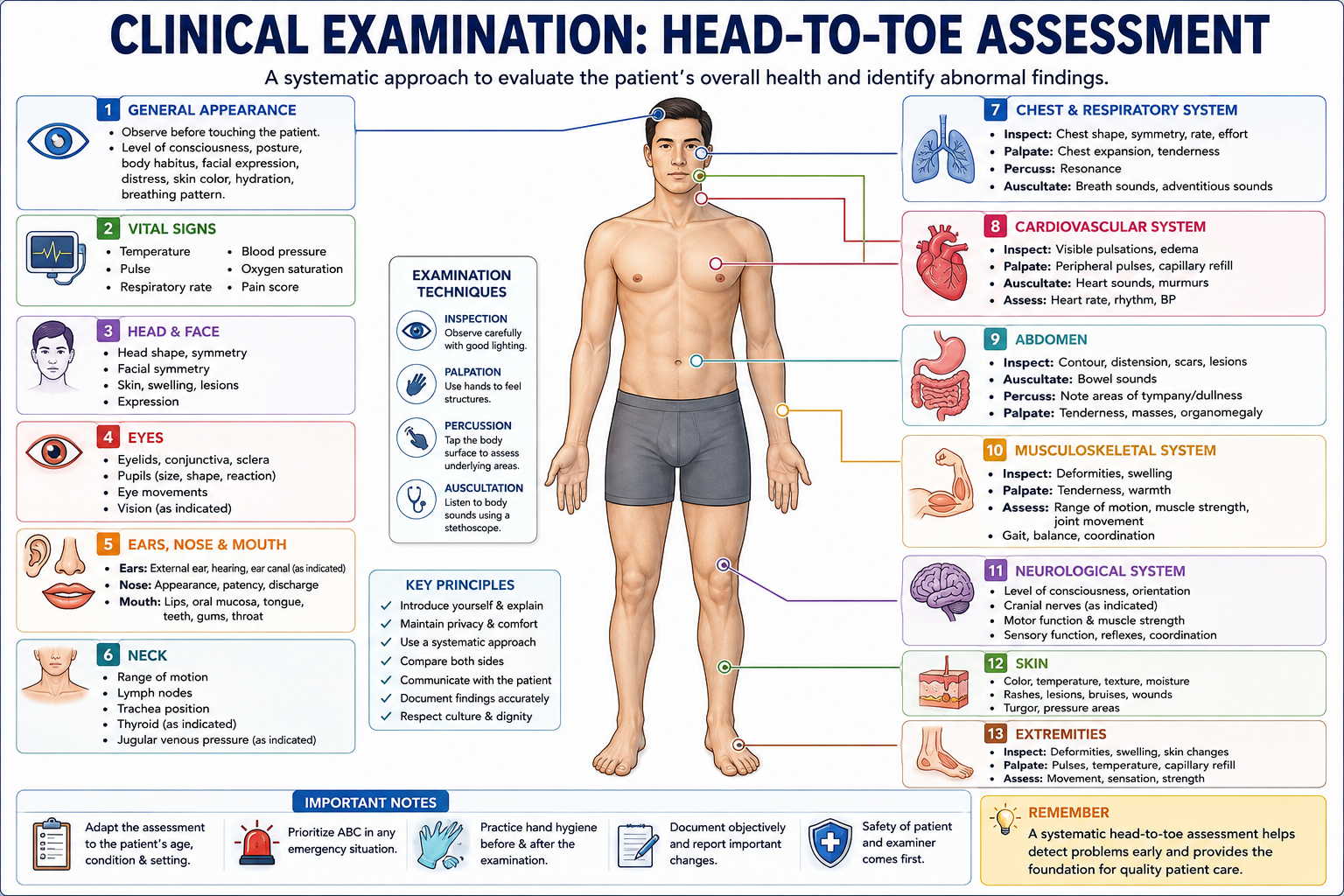
    A head-to-toe assessment is a systematic physical examination used by healthcare professionals to evaluate a patient’s overall health and identify abnormal findings.

    Instead of examining the patient randomly, the healthcare professional follows a structured sequence from the head down to the feet. This approach helps ensure that important body systems are assessed and that findings are documented consistently.

    For medical and nursing students, learning a systematic head-to-toe assessment is an important foundation for developing clinical examination skills.


    What Is a Head-to-Toe Assessment?

    A head-to-toe assessment is a comprehensive physical examination in which the healthcare professional evaluates the patient systematically.

    Depending on the clinical situation, the assessment may include:

    • General appearance
    • Vital signs
    • Head and face
    • Eyes and ears
    • Nose and mouth
    • Neck
    • Chest and respiratory system
    • Cardiovascular system
    • Abdomen
    • Musculoskeletal system
    • Neurological status
    • Skin
    • Extremities

    The exact examination can vary depending on the patient’s condition, age, symptoms, and clinical setting.


    1. Preparation Before the Examination

    Before beginning the assessment, proper preparation is important.

    Introduce Yourself

    Introduce yourself to the patient and explain what you are going to do.

    Confirm Patient Identity

    Use the appropriate patient-identification process according to the healthcare facility’s protocol.

    Explain the Examination

    Explain the purpose and general steps of the examination in language the patient can understand.

    Hand Hygiene

    Perform appropriate hand hygiene before and after patient contact.

    Privacy and Comfort

    Ensure appropriate privacy, lighting, positioning, and exposure only of the body areas necessary for the examination.


    2. General Appearance

    Begin by observing the patient before performing the detailed physical examination.

    Look at:

    • Level of consciousness
    • General appearance
    • Posture
    • Body position
    • Mobility
    • Facial expression
    • Skin color
    • Apparent distress
    • Breathing pattern
    • Communication ability

    This initial observation can provide important clues about the patient’s condition.


    3. Vital Signs

    Vital signs provide basic information about the patient’s physiological status.

    Depending on the clinical situation, assess:

    • Temperature
    • Pulse
    • Respiratory rate
    • Blood pressure
    • Oxygen saturation

    Pain assessment may also be included where appropriate.

    Abnormal vital signs should be interpreted together with the patient’s symptoms and other examination findings.


    4. Head and Face

    Examine the head and face for obvious abnormalities.

    Assess:

    • Head shape and symmetry
    • Facial symmetry
    • Facial movements
    • Skin condition
    • Swelling
    • Visible lesions

    Observe whether the patient appears comfortable and whether facial movements are symmetrical.


    5. Eyes

    The eye examination may include observation of:

    • Eyelids
    • Conjunctiva
    • Sclera
    • Pupils
    • Eye movements
    • Vision, when clinically indicated

    Pupillary Assessment

    Observe pupil size, symmetry, and reaction to light when appropriate.

    Abnormal pupil findings may require further neurological or ophthalmic assessment.


    6. Ears, Nose, and Mouth

    Ears

    Assess the external ears and, when indicated, hearing and the ear canal or tympanic membrane.

    Nose

    Observe:

    • Nasal appearance
    • Nasal passages
    • Discharge
    • Congestion
    • Breathing through the nose

    Mouth

    Assess:

    • Lips
    • Oral mucosa
    • Tongue
    • Teeth
    • Gums
    • Throat

    Look for abnormalities such as lesions, swelling, dryness, or changes in color.


    7. Neck

    Examine the neck for:

    • Range of motion
    • Swelling
    • Lymph nodes
    • Tracheal position
    • Visible abnormalities

    Depending on the clinical situation, assessment may also include examination of the thyroid or jugular venous pressure.


    8. Chest and Respiratory System

    The respiratory examination evaluates breathing and the condition of the lungs.

    Observe:

    • Respiratory rate
    • Respiratory effort
    • Chest movement
    • Chest symmetry
    • Use of accessory muscles
    • Signs of respiratory distress

    Depending on the clinical assessment, healthcare professionals may use:

    Inspection → Palpation → Percussion → Auscultation

    Auscultation allows assessment of breath sounds using a stethoscope.


    9. Cardiovascular Examination

    The cardiovascular examination focuses on the heart and circulation.

    Assess:

    • Heart rate
    • Heart rhythm
    • Peripheral pulses
    • Capillary refill when appropriate
    • Skin temperature
    • Peripheral edema
    • Heart sounds when indicated

    Auscultation of the heart may be performed using a stethoscope to assess heart sounds and identify possible abnormalities.


    10. Abdominal Examination

    The abdominal examination is generally performed systematically.

    A commonly taught sequence is:

    Inspection → Auscultation → Percussion → Palpation

    Inspection

    Look for:

    • Abdominal shape
    • Distension
    • Scars
    • Visible masses
    • Skin changes

    Auscultation

    Listen for bowel sounds and other relevant sounds when clinically indicated.

    Percussion

    Percussion can provide information about underlying structures and the presence of air, fluid, or solid tissue.

    Palpation

    Palpation can help assess:

    • Tenderness
    • Guarding
    • Masses
    • Organ enlargement

    The depth and extent of palpation should be appropriate to the clinical situation.


    11. Musculoskeletal Assessment

    The musculoskeletal examination evaluates movement, strength, and physical function.

    Assess:

    • Range of motion
    • Muscle strength
    • Joint movement
    • Swelling
    • Deformities
    • Gait when appropriate
    • Balance and coordination

    The assessment should be adapted according to the patient’s symptoms and mobility.


    12. Neurological Assessment

    A basic neurological examination may include assessment of:

    • Level of consciousness
    • Orientation
    • Speech
    • Motor function
    • Sensory function
    • Reflexes when indicated
    • Coordination
    • Balance
    • Cranial nerve function when appropriate

    The extent of neurological examination depends on the patient’s presentation.


    13. Skin Assessment

    Examine the skin for:

    • Color
    • Temperature
    • Moisture
    • Texture
    • Rashes
    • Wounds
    • Bruising
    • Pressure injuries
    • Swelling

    For patients with limited mobility, particular attention should be given to areas at risk of pressure injury.


    14. Extremities

    Examine the arms and legs for:

    • Symmetry
    • Movement
    • Muscle strength
    • Swelling
    • Skin changes
    • Peripheral pulses
    • Sensation
    • Capillary refill when appropriate

    Compare both sides of the body when clinically relevant.


    Inspection, Palpation, Percussion, and Auscultation

    Four fundamental techniques are commonly used during physical examination.

    Inspection

    Looking carefully at the patient and identifying visible findings.

    Palpation

    Using the hands to assess structures such as the skin, pulses, abdomen, lymph nodes, or joints.

    Percussion

    Tapping the body surface to help assess the characteristics of underlying structures.

    Auscultation

    Listening to body sounds, usually with a stethoscope.

    These techniques are combined differently depending on the body system being examined.


    Documentation of Findings

    Accurate documentation is an important part of clinical assessment.

    Record relevant findings clearly and objectively, including:

    • Vital signs
    • Positive findings
    • Significant negative findings
    • Patient symptoms
    • Examination results
    • Changes from previous assessments

    Avoid vague descriptions when a more objective description is possible.

    For example, instead of writing:

    “Patient looks unwell.”

    A more useful clinical description might document specific observable findings such as:

    “Patient appears pale and is breathing rapidly.”

    Documentation should follow the standards and terminology used by the healthcare institution.


    When Should the Assessment Be Modified?

    A complete head-to-toe assessment may not always be appropriate.

    For patients with an urgent or life-threatening problem, immediate priorities come first.

    For example, if a patient has severe breathing difficulty, the healthcare professional should prioritize airway, breathing, and circulation rather than delaying urgent care to complete a routine head-to-toe examination.

    The assessment should therefore be adapted to:

    • Patient condition
    • Presenting complaint
    • Age
    • Clinical setting
    • Level of consciousness
    • Emergency status
    • Relevant medical history

    Tips for Medical and Nursing Students

    1. Follow a Consistent Sequence

    Using the same general sequence helps reduce the chance of missing important findings.

    2. Practice With a Partner

    Practice examination techniques under appropriate supervision.

    3. Learn Normal Findings First

    Understanding what is normal makes it easier to recognize abnormalities.

    4. Communicate With the Patient

    Explain what you are doing and observe the patient’s response throughout the examination.

    5. Document Immediately

    Record important findings accurately according to your clinical documentation requirements.


    Conclusion

    A head-to-toe assessment provides a structured approach to evaluating a patient’s overall condition.

    By systematically assessing the general appearance, vital signs, head, eyes, ears, neck, respiratory system, cardiovascular system, abdomen, neurological system, musculoskeletal system, skin, and extremities, healthcare students can develop a strong foundation in clinical examination.

    The key is to combine a systematic approach with good communication, appropriate examination techniques, accurate documentation, and clinical judgment.

    A structured examination helps healthcare professionals recognize changes early and provide appropriate patient care.

  • Common Medical Emergencies and Their Management

    Common Medical Emergencies and Their Management

    Category: Emergency Medicine
    Tags: Medical Emergencies, First Aid, Emergency Management, CPR, Healthcare Professionals, Medical Students, Emergency Medicine

    Introduction

    Medical emergencies can occur suddenly and may require immediate assessment and treatment. Recognizing the warning signs of a serious condition and responding appropriately can help reduce complications and improve outcomes.

    Healthcare professionals and students should understand the basic principles of emergency assessment, prioritization, and initial management.

    This article provides an educational overview of common medical emergencies and the general principles used in their initial management. It is not a substitute for professional medical training or local emergency protocols.


    What Is a Medical Emergency?

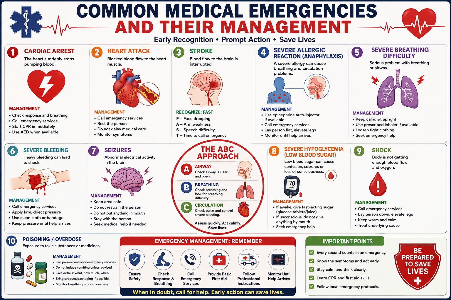
    A medical emergency is a condition that may pose an immediate threat to a person’s life or major bodily functions and requires prompt medical attention.

    Examples include:

    • Cardiac arrest
    • Heart attack
    • Stroke
    • Severe allergic reaction
    • Severe breathing difficulty
    • Severe bleeding
    • Seizures
    • Severe hypoglycemia
    • Shock
    • Poisoning or overdose

    Early recognition and appropriate action are important in all of these situations.


    1. Cardiac Arrest

    Cardiac arrest occurs when the heart suddenly stops effectively pumping blood.

    Warning Signs

    A person experiencing cardiac arrest may:

    • Become unresponsive
    • Stop breathing normally
    • Collapse suddenly

    Initial Management

    If cardiac arrest is suspected:

    1. Check that the scene is safe.
    2. Check whether the person responds.
    3. Assess breathing.
    4. Activate emergency medical services.
    5. Begin CPR if appropriate and follow local CPR guidance.
    6. Use an automated external defibrillator (AED) when available and follow its instructions.

    Rapid CPR and defibrillation when indicated can be critical.


    2. Heart Attack

    A heart attack, or myocardial infarction, occurs when blood flow to part of the heart muscle becomes blocked or severely reduced.

    Common Symptoms

    Symptoms can include:

    • Chest pressure, pain, or discomfort
    • Pain or discomfort spreading to the arm, shoulder, back, neck, or jaw
    • Shortness of breath
    • Sweating
    • Nausea
    • Unusual weakness or fatigue

    Symptoms can vary between individuals.

    Initial Management

    A suspected heart attack requires urgent medical evaluation.

    The person should stop exertion and seek emergency medical assistance. Do not delay emergency care while trying home remedies.

    Healthcare professionals may perform an ECG and other investigations to determine the cause of symptoms and guide treatment.


    3. Stroke

    A stroke occurs when blood flow to part of the brain is interrupted or when bleeding occurs within the brain.

    Recognizing a Stroke

    A useful approach is FAST:

    F — Face:
    Ask the person to smile. Is one side of the face drooping?

    A — Arms:
    Ask the person to raise both arms. Is one arm weak or drifting downward?

    S — Speech:
    Is speech unclear, unusual, or difficult to understand?

    T — Time:
    Treat the situation as an emergency and seek medical help immediately.

    Other symptoms can include sudden vision problems, dizziness, difficulty walking, confusion, or a severe headache.

    Initial Management

    Call emergency medical services immediately.

    Note the time when symptoms were first noticed, if known. Do not give food, drink, or medication unless instructed by a qualified healthcare professional.

    4. Severe Allergic Reaction

    A severe allergic reaction, known as anaphylaxis, can develop rapidly and may affect breathing and circulation.

    Possible Symptoms

    Symptoms may include:

    • Difficulty breathing
    • Swelling of the lips, tongue, or throat
    • Wheezing
    • Widespread hives
    • Dizziness or fainting
    • Low blood pressure
    • Rapid deterioration

    Initial Management

    Anaphylaxis requires immediate emergency treatment.

    If the person has a prescribed epinephrine/adrenaline auto-injector, it should be used according to their emergency action plan and device instructions.

    Emergency medical services should be contacted immediately, even if symptoms begin to improve.


    5. Severe Breathing Difficulty

    Breathing difficulty can result from conditions such as asthma, severe allergic reactions, pneumonia, airway obstruction, or heart problems.

    Warning Signs

    Seek urgent medical attention when a person has:

    • Severe difficulty breathing
    • Blue or gray lips or skin
    • Inability to speak normally because of breathlessness
    • Severe wheezing or noisy breathing
    • Confusion or reduced consciousness
    • Rapid deterioration

    Initial Management

    Keep the person in a position that makes breathing easier and seek emergency medical assistance.

    If the person has a prescribed rescue medication, such as an asthma inhaler, help them use it according to their existing medical instructions.


    6. Severe Bleeding

    Severe bleeding can become life-threatening if blood loss is significant.

    Signs

    Possible signs include:

    • Continuous or heavy bleeding
    • Blood rapidly soaking through clothing or dressings
    • Weakness or dizziness
    • Pale or clammy skin
    • Loss of consciousness

    Initial Management

    If it is safe to do so:

    1. Call emergency medical services.
    2. Apply firm, direct pressure to the wound using appropriate material.
    3. Continue pressure and do not repeatedly remove dressings to check the wound.
    4. Follow emergency responder instructions.

    For life-threatening limb bleeding, trained responders may use a tourniquet according to appropriate emergency protocols.


    7. Seizures

    A seizure can cause sudden changes in awareness, movement, sensation, or behavior.

    During a generalized convulsive seizure, the person may become unconscious and experience uncontrolled movements.

    What To Do

    During a seizure:

    • Keep the surrounding area safe.
    • Move dangerous objects away.
    • Protect the person’s head if possible.
    • Do not restrain the person.
    • Do not put anything in their mouth.
    • Do not give food or drink until they are fully alert.
    • After the seizure, monitor their breathing and stay with them.

    Seek emergency medical assistance when the seizure is prolonged, repeats without recovery, occurs in water, causes serious injury, involves breathing difficulties, or occurs in someone who is pregnant or has another significant medical concern.


    8. Severe Hypoglycemia

    Hypoglycemia means that blood glucose is too low. It can occur particularly in people using certain diabetes medications.

    Possible Symptoms

    Early symptoms may include:

    • Sweating
    • Shaking
    • Hunger
    • Dizziness
    • Irritability
    • Confusion
    • Weakness

    Severe hypoglycemia can cause seizures or loss of consciousness.

    Initial Management

    If the person is awake and able to swallow safely, appropriate fast-acting carbohydrate may be given according to their diabetes care plan.

    If the person is unconscious or unable to swallow safely, do not give food or drink by mouth. Seek emergency medical assistance and follow the person’s emergency diabetes plan if available.


    9. Shock

    Shock occurs when the body’s tissues do not receive adequate blood flow and oxygen.

    It can result from severe bleeding, infection, major injury, severe allergic reactions, or other serious conditions.

    Possible Signs

    A person in shock may have:

    • Pale or clammy skin
    • Rapid breathing
    • Weakness
    • Dizziness
    • Confusion
    • Rapid pulse
    • Reduced consciousness

    Initial Management

    Shock is a medical emergency.

    Call emergency medical services and address the underlying cause if trained to do so. Keep the person warm and monitor their breathing and responsiveness while waiting for professional help.


    10. Poisoning and Overdose

    Poisoning can occur after exposure to medicines, chemicals, household products, or other toxic substances.

    Initial Management

    Do not make the person vomit unless specifically instructed by a poison control professional or healthcare provider.

    Seek emergency medical advice and provide information about:

    • What substance was involved
    • Approximately how much was involved
    • When the exposure occurred
    • The person’s age and symptoms
    • The packaging or product information, if available

    If the person is unconscious, having seizures, or having difficulty breathing, seek emergency medical assistance immediately.


    The ABC Approach to Emergency Assessment

    Healthcare professionals commonly use structured approaches to rapidly assess critically ill patients.

    A basic framework is:

    A — Airway

    Check whether the airway is open and whether there is an obstruction.

    B — Breathing

    Assess breathing and look for signs of respiratory distress.

    C — Circulation

    Assess circulation and look for severe bleeding or signs of poor perfusion.

    In professional emergency care, assessment continues with additional steps such as neurological evaluation and exposure/environmental assessment.


    Why Early Recognition Matters

    Many emergencies become more difficult to manage as time passes.

    Recognizing warning signs early can help ensure that appropriate medical assistance is requested without unnecessary delay.

    For healthcare students, learning emergency medicine should include:

    • Recognizing life-threatening conditions
    • Understanding basic emergency assessment
    • Learning CPR and first-aid skills through appropriate training
    • Understanding when to activate emergency services
    • Following local clinical protocols
    • Communicating clearly with emergency teams

    Emergency Management: Remember

    When faced with a suspected medical emergency:

    1. Stay calm and assess the situation.
    2. Make sure the environment is safe.
    3. Check responsiveness and breathing.
    4. Call emergency medical services when appropriate.
    5. Provide basic first aid within your level of training.
    6. Follow instructions from emergency professionals.
    7. Continue monitoring the person until help arrives.

    Conclusion

    Medical emergencies require rapid recognition, appropriate prioritization, and timely professional care.

    Understanding conditions such as cardiac arrest, heart attack, stroke, anaphylaxis, severe breathing difficulty, major bleeding, seizures, hypoglycemia, shock, and poisoning provides an important foundation for healthcare students.

    However, reading about emergency management is not a replacement for practical training. Students and healthcare professionals should learn emergency procedures through recognized training programs and follow the protocols used by their healthcare institution and local emergency services.

    Early recognition, appropriate first response, and rapid access to professional medical care can make a critical difference in an emergency.

  • How ECG Works: A Beginner’s Guide

    How ECG Works: A Beginner’s Guide

    Category: Human Anatomy
    Tags: ECG, Electrocardiogram, Heart, Cardiology, Heart Rhythm, Medical Students, Cardiovascular System

    Introduction

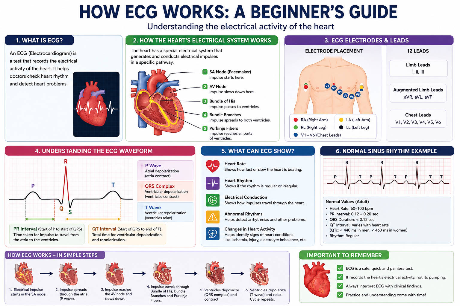
    An electrocardiogram (ECG or EKG) is a simple and widely used medical test that records the electrical activity of the heart. It helps healthcare professionals understand how the heart is beating and can provide important information about heart rhythm and electrical conduction….

    For medical and healthcare students, learning how an ECG works is an important foundation for understanding cardiac physiology and common heart conditions.

    In this beginner’s guide, we will explain what an ECG is, how it records the heart’s electrical activity, what the main ECG components represent, and how healthcare professionals use ECGs in clinical practice.

    What Is an ECG?

    An ECG is a test that records the electrical signals produced by the heart.

    Every heartbeat is controlled by electrical impulses. These impulses cause the heart muscle to contract in a coordinated manner, allowing blood to be pumped through the heart and into the circulation.

    An ECG detects these electrical changes through electrodes placed on the skin and displays them as a waveform.

    The test is:

    • Non-invasive
    • Usually painless
    • Relatively quick
    • Commonly performed in hospitals and clinics

    How Does the Heart Produce Electrical Signals?

    The heartbeat is controlled by a specialized electrical conduction system.

    The electrical impulse normally begins in the sinoatrial (SA) node, located in the right atrium.

    The signal then travels through the heart in a coordinated sequence:

    SA Node → Atria → AV Node → Bundle of His → Bundle Branches → Purkinje Fibers → Ventricles

    This electrical activity causes the atria and ventricles to contract in the appropriate sequence.

    How Does an ECG Machine Detect the Signal?

    The ECG machine does not directly measure the heart’s mechanical pumping action.

    Instead, it detects changes in electrical potential on the body’s surface.

    Small adhesive electrodes are placed on specific locations on the patient’s chest and limbs. These electrodes detect electrical signals generated by the heart.

    The ECG machine processes these signals and displays them as waveforms.

    A healthcare professional can then examine the waveform to assess the heart’s electrical activity.


    ECG Electrodes

    A standard 12-lead ECG uses 10 physical electrodes to create 12 different electrical views, or leads, of the heart.

    The electrodes are generally placed on:

    Limb Electrodes

    Four electrodes are placed on the limbs:

    • Right arm
    • Left arm
    • Right leg
    • Left leg

    Chest Electrodes

    Six chest electrodes are placed at specific locations across the chest:

    • V1
    • V2
    • V3
    • V4
    • V5
    • V6

    Together, these electrodes provide different views of the heart’s electrical activity.

    What Are ECG Leads?

    An ECG lead represents a particular electrical viewpoint of the heart.

    The standard 12 leads are:

    Limb Leads

    • Lead I
    • Lead II
    • Lead III

    Augmented Limb Leads

    • aVR
    • aVL
    • aVF

    Chest Leads

    • V1
    • V2
    • V3
    • V4
    • V5
    • V6

    Each lead provides information about electrical activity from a different direction.

    This allows healthcare professionals to examine different regions of the heart.


    Understanding the ECG Waveform

    A normal ECG waveform contains several important components.

    The most commonly discussed are:

    • P wave
    • QRS complex
    • T wave
    • PR interval
    • QT interval

    Let’s understand them one by one.


    P Wave

    The P wave represents atrial depolarization.

    In simple terms, it reflects the electrical activation of the atria before they contract.

    A healthcare professional can examine the P wave to assess aspects of atrial electrical activity.


    QRS Complex

    The QRS complex represents ventricular depolarization.

    It is usually the most prominent part of the ECG waveform.

    The QRS complex reflects the electrical activation of the ventricles, which precedes ventricular contraction.


    T Wave

    The T wave represents ventricular repolarization.

    It reflects the electrical recovery of the ventricles after their activation.

    The P wave, QRS complex, and T wave together provide a basic framework for understanding the electrical cycle of the heart.


    PR Interval

    The PR interval represents the time between the beginning of atrial depolarization and the beginning of ventricular depolarization.

    It includes the time required for the electrical impulse to travel through the atria and the atrioventricular conduction system.


    QT Interval

    The QT interval represents the duration of ventricular electrical activity, from ventricular depolarization through ventricular repolarization.

    The QT interval is important in clinical ECG interpretation because its duration can vary with heart rate and other factors.


    What Can an ECG Show?

    An ECG can provide information about several aspects of cardiac electrical activity.

    Healthcare professionals may use it to assess:

    Heart Rate

    The ECG can help determine how quickly the heart is beating.

    Heart Rhythm

    It can help identify whether the heartbeat follows a regular rhythm.

    Electrical Conduction

    An ECG can provide information about how electrical impulses travel through the heart.

    Abnormal Rhythms

    ECG recordings can help identify various cardiac arrhythmias.

    Changes in Cardiac Electrical Activity

    Certain heart conditions can produce characteristic changes in ECG patterns.

    An ECG is only one part of a clinical assessment. Healthcare professionals interpret ECG findings together with symptoms, medical history, physical examination, and other investigations when necessary.


    Why Is ECG Important for Medical Students?

    ECG interpretation combines concepts from anatomy, physiology, and clinical medicine.

    A good starting point for beginners is to understand:

    1. How the heart’s electrical system works.
    2. How electrical impulses travel through the heart.
    3. What the P wave represents.
    4. What the QRS complex represents.
    5. What the T wave represents.
    6. How the 12 ECG leads provide different views of the heart.
    7. How heart rate and rhythm are assessed.

    Once these concepts are clear, students can gradually move toward more advanced ECG interpretation.


    Basic ECG Learning Approach

    If you are learning ECG for the first time, avoid trying to memorize every abnormal pattern immediately.

    A useful learning sequence is:

    Step 1: Understand Cardiac Anatomy

    Learn the four chambers of the heart and the major blood vessels.

    Step 2: Learn Cardiac Electrical Conduction

    Understand the pathway:

    SA Node → AV Node → Bundle of His → Bundle Branches → Purkinje Fibers

    Step 3: Learn the Normal ECG

    Understand the:

    P Wave → QRS Complex → T Wave

    Step 4: Learn Heart Rate and Rhythm

    Practice identifying regular and irregular rhythms.

    Step 5: Study Common Abnormalities

    After understanding a normal ECG, move on to common abnormalities and arrhythmias under appropriate clinical supervision or educational resources.


    ECG vs. Echocardiogram

    ECG and echocardiography are different tests.

    ECGEchocardiogram
    Records electrical activityUses ultrasound to create images of the heart
    Shows electrical rhythm and conductionShows heart structure and movement
    Uses electrodesUses an ultrasound transducer
    Produces waveformsProduces images/video

    Understanding this difference is useful for medical students because both tests are commonly used in cardiovascular medicine but provide different types of information.


    Conclusion

    An ECG is an important tool for recording and evaluating the electrical activity of the heart.

    For beginners, the most important concepts are the cardiac conduction system, ECG electrodes and leads, P wave, QRS complex, T wave, and basic intervals.

    Once these fundamentals are understood, students can progress to more advanced topics such as rhythm interpretation, conduction abnormalities, ischemic changes, and other clinical ECG patterns.

    Learning the normal ECG is the first step toward understanding abnormal cardiac rhythms and electrical activity.

  • Understanding the Cardiovascular System

    Understanding the Cardiovascular System

    Category: Medical Education
    Tags: Cardiovascular System, Human Anatomy, Heart, Blood Circulation, Medical Students

    Introduction

    The cardiovascular system is one of the most important systems in the human body. It is responsible for circulating blood throughout the body, delivering oxygen and nutrients to tissues, and transporting waste products away from cells….

    For medical and healthcare students, understanding the cardiovascular system is essential because it forms the foundation for studying conditions such as hypertension, heart failure, coronary artery disease, and other cardiovascular disorders.

    In this article, we will explore the major components of the cardiovascular system, how blood circulates through the body, and some important concepts every beginner should understand.

    What Is the Cardiovascular System?

    The cardiovascular system, also called the circulatory system, consists mainly of three components:

    1. Heart
    2. Blood vessels
    3. Blood

    Together, these components work continuously to transport substances throughout the body.

    The heart acts as the pump, blood vessels provide the transportation network, and blood carries oxygen, nutrients, hormones, and waste products.

    The Heart

    The heart is a muscular organ located in the chest between the lungs. Its primary function is to pump blood throughout the body.

    The adult human heart has four chambers:

    • Right atrium
    • Right ventricle
    • Left atrium
    • Left ventricle

    Right Atrium

    The right atrium receives oxygen-poor blood returning from the body through the major veins.

    Right Ventricle

    The right ventricle pumps oxygen-poor blood toward the lungs through the pulmonary arteries.

    Left Atrium

    The left atrium receives oxygen-rich blood returning from the lungs through the pulmonary veins.

    Left Ventricle

    The left ventricle pumps oxygen-rich blood into the systemic circulation through the aorta.

    The left ventricle has a relatively thick muscular wall because it must generate enough pressure to circulate blood throughout the body.

    Heart Valves

    Heart valves help ensure that blood flows in the correct direction.

    The four major valves are:

    • Tricuspid valve
    • Pulmonary valve
    • Mitral valve
    • Aortic valve

    These valves open and close according to pressure changes within the heart.

    For example, the tricuspid valve allows blood to move from the right atrium to the right ventricle while helping prevent backward flow.

    How Blood Circulates Through the Heart

    Understanding the pathway of blood through the heart is an important foundation for studying cardiovascular anatomy.

    The simplified pathway is:

    Body → Right Atrium → Right Ventricle → Lungs → Left Atrium → Left Ventricle → Body

    More specifically:

    1. Oxygen-poor blood returns from the body.
    2. Blood enters the right atrium.
    3. It moves into the right ventricle.
    4. The right ventricle pumps blood to the lungs.
    5. Blood receives oxygen in the lungs.
    6. Oxygen-rich blood returns to the left atrium.
    7. It moves into the left ventricle.
    8. The left ventricle pumps blood into the aorta.
    9. Blood is distributed throughout the body.

    Blood Vessels

    Blood vessels form an extensive network throughout the body.

    There are three primary types:

    Arteries

    Arteries generally carry blood away from the heart.

    Most arteries carry oxygen-rich blood. However, the pulmonary arteries are an important exception because they carry oxygen-poor blood from the heart to the lungs.

    Veins

    Veins generally carry blood toward the heart.

    Most veins carry oxygen-poor blood. The pulmonary veins are an important exception because they carry oxygen-rich blood from the lungs to the heart.

    Capillaries

    Capillaries are very small blood vessels that connect the arterial and venous sides of circulation.

    They are important sites for the exchange of:

    • Oxygen
    • Carbon dioxide
    • Nutrients
    • Waste products
    • Other substances between blood and tissues

    Pulmonary and Systemic Circulation

    The cardiovascular system can be broadly divided into two major circulation pathways.

    Pulmonary Circulation

    Pulmonary circulation carries blood between the heart and lungs.

    Its primary purpose is to allow blood to exchange carbon dioxide for oxygen.

    Right ventricle → Pulmonary arteries → Lungs → Pulmonary veins → Left atrium

    Systemic Circulation

    Systemic circulation carries oxygen-rich blood from the heart to the body’s tissues and returns oxygen-poor blood back to the heart.

    Left ventricle → Aorta → Body tissues → Veins → Right atrium

    Understanding the difference between pulmonary and systemic circulation is essential for learning cardiovascular physiology.

    What Is Blood Pressure?

    Blood pressure is the force exerted by circulating blood against the walls of blood vessels.

    It is commonly represented using two numbers:

    • Systolic pressure — pressure when the heart contracts.
    • Diastolic pressure — pressure when the heart relaxes between beats.

    Blood pressure is an important clinical measurement because abnormal blood pressure can be associated with various cardiovascular conditions.

    Heart Rate

    Heart rate refers to the number of times the heart beats per minute.

    The heart’s rhythm is coordinated by its electrical conduction system.

    Important structures include:

    • Sinoatrial (SA) node
    • Atrioventricular (AV) node
    • Bundle of His
    • Bundle branches
    • Purkinje fibers

    The SA node normally acts as the heart’s natural pacemaker and initiates electrical impulses that help coordinate the heartbeat.

    Coronary Circulation

    The heart itself requires oxygen and nutrients to function. These are supplied by the coronary arteries.

    Coronary arteries arise from the aorta and supply blood to the heart muscle.

    If blood flow through a coronary artery becomes significantly restricted, the heart muscle may not receive enough oxygen.

    This is an important concept when studying conditions such as coronary artery disease and myocardial infarction.

    Why Is the Cardiovascular System Important?

    The cardiovascular system performs several essential functions, including:

    • Delivering oxygen to tissues
    • Transporting nutrients
    • Removing metabolic waste
    • Transporting hormones
    • Helping regulate body temperature
    • Supporting immune responses
    • Maintaining circulation and tissue perfusion

    Because virtually every organ depends on adequate blood supply, cardiovascular health is essential for overall health.

    Tips for Medical Students

    The cardiovascular system can initially seem complicated because it combines anatomy, physiology, and clinical concepts.

    Here are some useful study strategies:

    1. Memorize the Blood Flow Pathway

    Start by learning:

    Body → Right Heart → Lungs → Left Heart → Body

    Once this pathway is clear, many cardiovascular concepts become easier.

    2. Study the Heart With Diagrams

    Use labeled diagrams to identify the chambers, valves, major vessels, and coronary arteries.

    3. Understand Before Memorizing

    Rather than simply memorizing the names of structures, understand what each structure does and where blood moves next.

    4. Connect Anatomy With Clinical Medicine

    After learning normal cardiovascular anatomy, relate it to common conditions such as hypertension, heart failure, arrhythmias, and coronary artery disease.

    Conclusion

    The cardiovascular system is a complex but highly organized network responsible for maintaining continuous blood circulation throughout the body.

    Understanding the heart chambers, valves, blood vessels, circulation pathways, blood pressure, and electrical conduction system provides a strong foundation for further study in physiology and clinical medicine.

    For beginners, the best approach is to learn the system step by step, beginning with the structure of the heart and the pathway of blood before moving into more advanced cardiovascular physiology and disease processes.

  • Human Anatomy Basics for Medical Students: A Beginner’s Guide

    Human Anatomy Basics for Medical Students: A Beginner’s Guide

    Human anatomy is one of the most important subjects for every medical student. It helps us understand the structure of the human body and forms the foundation for learning physiology, pathology, surgery, and clinical medicine.

    Whether you are studying MBBS, Nursing, Pharmacy, or Physiotherapy, learning anatomy is the first step toward becoming a healthcare professional..

    In this guide, we’ll explore the basics of human anatomy, the major body systems, and useful study tips for beginners.

    What Is Human Anatomy?

    Human anatomy is the branch of science that studies the structure of the human body. It focuses on organs, bones, muscles, tissues, and how they are arranged inside the body.

    Anatomy is usually studied together with physiology, which explains how these body parts work.

    Why Is Anatomy Important?

    • Understand body structure
    • Diagnose diseases
    • Perform surgeries safely
    • Interpret X-rays and MRI scans
    • Improve patient care
    • Learn medical terminology

    Major Organ Systems

    • Skeletal System

    Supports the body, protects organs, stores minerals, and produces blood cells.

    • Muscular System

    Responsible for movement, posture, and heat production.

    • Nervous System

    Controls the brain, spinal cord, and nerves.

    • Cardiovascular System

    Circulates blood throughout the body.

    • Respiratory System
    • Digestive System
    • Urinary System
    • Endocrine System
    • Reproductive System
    • Integumentary System
    TermMeaning
    SuperiorTowards the head
    InferiorTowards the feet
    AnteriorFront
    PosteriorBack
    MedialTowards the middle

    Frequently Asked Questions

    Is anatomy difficult?

    It can be challenging at first, but regular practice and revision make it easier.

    How many bones are in the adult body?

    An adult human has 206 bones.

    Conclusion

    Human anatomy is the foundation of medical education. By understanding the body’s structure and organ systems, students develop the knowledge required for diagnosis, treatment, and patient care. Building a strong understanding of anatomy early will help throughout your medical career.